Your eye exam photos could reveal your Alzheimer’s risk years before symptoms start
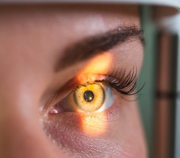
The retina at the back of your eye is a thin layer of tissue, but it has a direct connection to your brain through the optic nerve. That link has long made it an object of scientific curiosity. Now, a study from the University of Florida suggests that everyday photos taken during routine eye exams could be used to flag who is at risk of developing Alzheimer's disease, often years or even decades before any symptoms appear.
Researchers trained an AI model on retinal photographs from more than 40,000 patients in a UK patient database. The model learned to pick out subtle features in the eye that correspond to known Alzheimer's risk factors, including high blood pressure, smoking, alcohol use, and even insomnia. The results were published on June 16 in the Journal of Alzheimer's Disease.
The lead researcher, Ruogu Fang, a professor of biomedical engineering at UF, framed the finding simply: "We know that Alzheimer's disease develops over decades, but most of the diagnostic tools focus on late-stage pathology when it is too late to intervene. By looking at novel biomarkers, like retinal health, we offer new opportunities to identify patients at risk, offer appropriate tests and encourage them to develop healthy lifestyles to mitigate their risk."
How does it work?
Retinal photographs are already a standard part of eye care for millions of people. Anyone being monitored for diabetes, glaucoma, or cataracts will have had multiple retinal scans over the years. Even a basic glasses prescription check can include one. That makes the images widely available and cheap to collect compared to brain MRIs or other specialist scans.
The AI model analyzed those photographs and identified specific structures in the eye linked to Alzheimer's risk, particularly the arteries and the optic nerve. From those images alone, the model could accurately predict:
- Biological traits like sex and blood pressure
- Lifestyle factors including smoking and alcohol consumption
- Sleep-related issues such as insomnia
Many of these details already exist somewhere in a patient's medical records, but those records are often incomplete. Self-reported data on smoking or drinking is notoriously unreliable. Retinal images offer something different: a physical record of cumulative damage built up over time, which can vary between people who share similar risk profiles on paper.
Fang described retinal imaging as "an integrated biological sensor of cumulative risk" rather than just another way to ask patients about their habits.
Why does it matter?
The biggest problem with Alzheimer's diagnosis right now is timing. By the time a patient shows noticeable cognitive decline, significant brain damage has already occurred. Treatments and lifestyle interventions have much more room to work earlier in that process.
Fang's team has already published research showing that retinal photographs can detect active Alzheimer's disease. This new study goes further by targeting risk factors in people who have not yet developed the condition. That opens up the possibility of identifying at-risk patients early enough to act, whether that means:
- Encouraging protective lifestyle changes
- Starting certain medications earlier
- Referring patients for cognitive training programs
- Triggering more targeted follow-up testing
Seowung Leem, a doctoral student at UF and the paper's first author, put it this way: "With the assistance of AI, we are now able to identify subtle retinal variations that were formerly overlooked across thousands of subjects, which may function as reliable indicators of future disease risk."
The context
Alzheimer's affects tens of millions of people worldwide, and that number is expected to rise sharply as populations age. Despite decades of research, there is still no cure, and most approved treatments only slow progression rather than stop it. That has pushed researchers to look harder at prevention and early detection.
The idea of using the eye as a window into brain health is not new, but it has gained serious momentum in recent years. Researchers have explored retinal imaging as a way to detect signs of multiple sclerosis, Parkinson's disease, and cardiovascular risk. The availability of large patient datasets and more capable AI tools has made it possible to find patterns in retinal images that human clinicians would miss.
This study used data from the UK Biobank, one of the largest and most detailed health databases in the world, which has become a key resource for this kind of population-scale research. The work was supported in part by the National Science Foundation, and the team included researchers from UF's McKnight Brain Institute and Meta.
The practical implication is straightforward: a test that millions of people already take, for completely different reasons, could also serve as an early warning system for one of the most feared diseases of aging. No extra equipment required.
Latest @ 
Abu Dhabi mental health network teams up with speech therapy clinic to tackle digital wellbeing
Abu Dhabi partners with Eli Lilly to tackle obesity and Alzheimer's with AI
Abu Dhabi launches first genomics-driven Alzheimer's trial using national DNA database
Abu Dhabi Biobank partners with BioTwin to bring AI 'virtual human twin' technology to the UAE
Abu Dhabi and Boehringer Ingelheim team up to advance precision medicine research
💡Did you know?
You can take your DHArab experience to the next level with our Premium Membership.👉 Click here to learn more
🛠️Featured tool
 Easy-Peasy
An all-in-one AI tool offering the ability to build no-code AI Bots, create articles & social media posts, convert text into natural speech in 40+ languages, create and edit images, generate videos, and more.
👉 Click here to learn more
