

AI reads the “geography” of pancreatic tumors to predict cancer recurrence
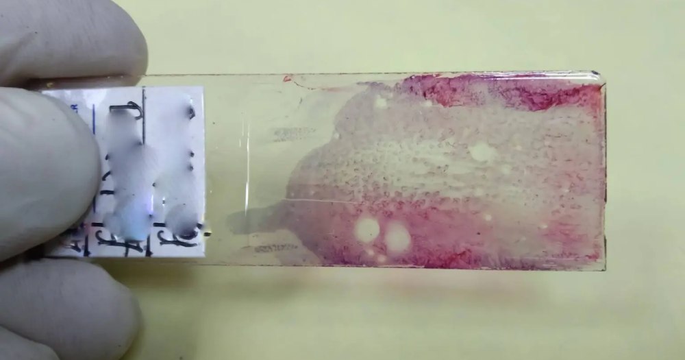
It's not the amount of cancer left after treatment that best predicts whether pancreatic cancer will return. It's the shape of it. That finding, published in Clinical Cancer Research, could shift how pathologists and oncologists interpret post-treatment tissue slides.
Researchers at Mayo Clinic trained an AI tool to analyze standard pathology slides from 203 patients with pancreatic ductal adenocarcinoma who had received chemotherapy before surgery but showed only a limited response. Rather than counting residual cancer cells, the tool mapped how cancer tissue and surrounding stroma were spatially arranged, measuring fragmentation, boundary patterns, and the degree to which the two tissue types were intermixed.
Patients whose tissue showed a more fragmented, intermixed pattern experienced earlier recurrence. Two spatial models identified high-risk patients with 71% and more than double the adjusted recurrence risk, respectively, even after accounting for stage, lymph node status, and other standard clinical factors. Standard measures alone, including how much tumor remained, did not reliably separate the groups.
How does it work?
The team combined an AI-enabled digital pathology platform with methods adapted from landscape ecology to analyze routine hematoxylin and eosin slides already produced as part of standard care. The AI identified cancer and stromal regions, then measured spatial features including tissue shape, fragmentation, and intermixing. No additional tissue tests are required. The analysis works on slides that already exist.
Why does it matter?
Pancreatic cancer has one of the lowest survival rates of any major cancer, and recurrence after surgery remains a significant clinical problem. Current pathology assessments, as senior author Ryan Carr notes, tell clinicians how much tumor is left, not what the biology of that remaining tissue suggests about future behavior. This approach adds a layer of spatial information that standard assessment misses. The study also found that high-risk spatial patterns contained fewer immune cells within the tumor itself, pointing to a link between tissue architecture and immune exclusion.
The context
Across the GCC, pancreatic cancer is among the harder malignancies to catch early, and access to advanced pathology interpretation varies considerably between major cancer centers and smaller facilities. AI-assisted digital pathology offers a practical path forward because it works on existing infrastructure. For health systems in Saudi Arabia and the UAE investing in AI-enabled diagnostics under Vision 2030 and national health transformation programs, this kind of tool, built on routine slides rather than expensive new tests, is exactly the type of scalable application that fits the regional agenda. The Mayo team cautions that prospective studies are still needed before the approach enters clinical decision-making.
Latest @
Tunisia bets on telehealth as it prepares to host Telehealth Connect 2026
Wearable AI system developed by Qatar medical students flags cardiac risk outside hospital
GLP-1 consultations surge 60% at Valeo Health as UAE weight management shifts toward continuous care
Abu Dhabi sends first health mission to China, targeting AI, cancer care and new drug development
UAE pharmaceutical chief meets Moderna co-founder in New York to explore manufacturing and R&D ties
💡Did you know?
You can take your DHArab experience to the next level with our Premium Membership.👉 Click here to learn more
🛠️Featured tool
 Easy-Peasy
An all-in-one AI tool offering the ability to build no-code AI Bots, create articles & social media posts, convert text into natural speech in 40+ languages, create and edit images, generate videos, and more.
👉 Click here to learn more
