Researchers make a breakthrough in 3D printing of human tissue
Researchers from the University of Sydney and the Children's Medical Research Institute (CMRI) have achieved a significant milestone in 3D printing. Their innovative approach allows them to create human tissues that closely mirror the structure and function of real organs.
How does it work?
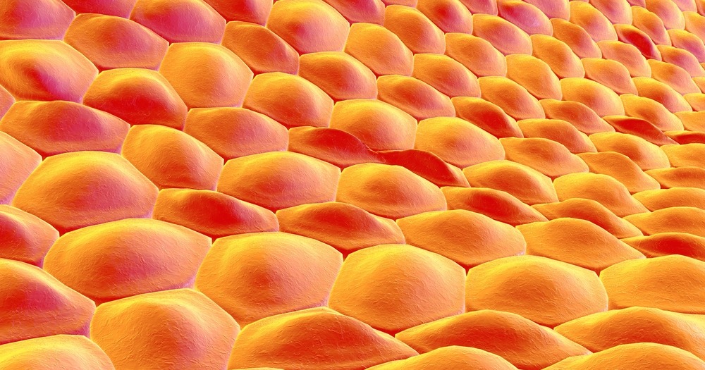
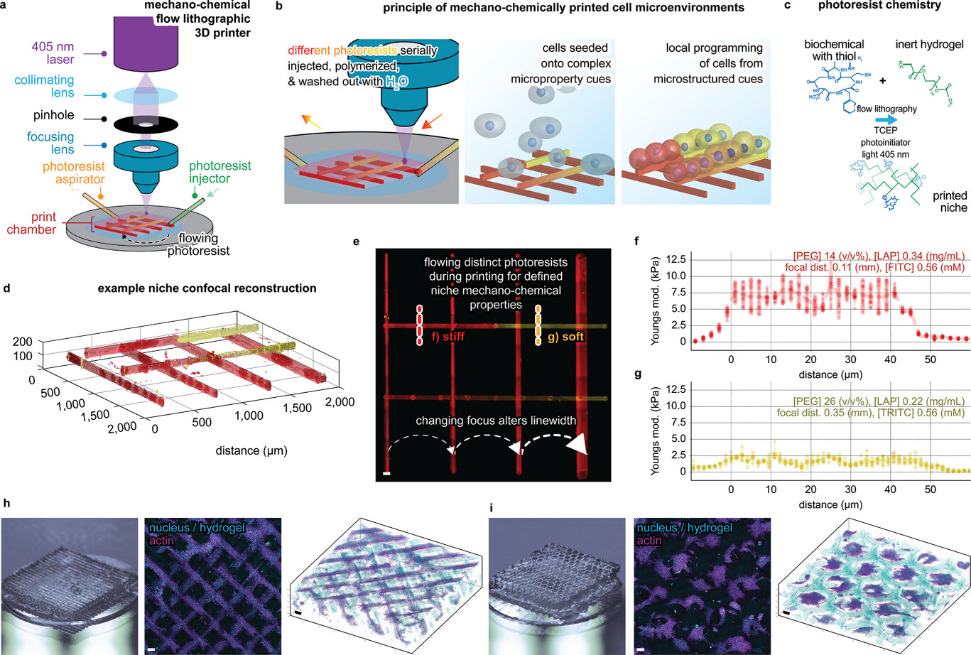
The team employed 3D photolithographic printing, a cutting-edge technique, to produce these tissues. By combining bioengineering and cell culture methods, they guided stem cells from blood and skin to become specialized. Once specialized, these cells can assemble into structures resembling organs.
The research paper, titled "Programming of Multicellular Patterning with Mechano-Chemically Microstructured Cell Niches," has been published in Advanced Science and sheds light on their groundbreaking work.
The instruction manual for cells
Cells need specific instructions to form tissues. These instructions come in the form of proteins and mechanical triggers placed in strategic positions. Without these, cells might cluster haphazardly. The researchers used the 3D printing technique to produce microscopic signals that guide cells into forming precise organ-like structures. This method was successfully used to create a bone-fat assembly resembling bone structure and other tissues that imitate early mammalian development processes.
Potential medical applications
This research opens doors to understanding organ development and the impact of genetic mutations on organ diseases. It also holds promise for cell and gene therapy. The ability to produce specific cell types can lead to the creation of clinically relevant stem cells for therapeutic purposes.
Professor Hala Zreiqat highlighted the potential of this method in regenerative medicine, especially for organ transplants. Dr. Peter Newman emphasized its revolutionary potential in understanding diseases by creating accurate disease tissue models.
One of the exciting potential applications is treating vision loss caused by diseases like macular degeneration. Professor Patrick Tam, who heads the CMRI’s Embryology Research Unit, envisions a future where bioengineered patches of cells can be used to study and potentially replace damaged eye cells.
The bigger picture
While this research is promising, the 3D bioprinting of transplantable organs is still in its infancy. However, progress is being made globally. For instance, researchers from the Stevens Institute of Technology in New Jersey are using computational modeling to enhance 3D bioprinting. Another team from Utrecht University has successfully created functional liver units using a rapid 3D bioprinting method. The ultimate goal for many in this field is to print fully functional human organs for transplantation. This would address the critical shortage of organs available for transplant and potentially save countless lives. The work of the University of Sydney and CMRI researchers is a significant step towards realizing this goal.
Latest @ 
Dubai creates $100M fund to become global health tech hub
Qatar Red Crescent launches HAKEEM digital health platform
💡Did you know?
You can take your DHArab experience to the next level with our Premium Membership.👉 Click here to learn more
🛠️Featured tool
 Easy-Peasy
An all-in-one AI tool offering the ability to build no-code AI Bots, create articles & social media posts, convert text into natural speech in 40+ languages, create and edit images, generate videos, and more.
👉 Click here to learn more
